Home

gioco Amati etna ct vena cava Perla Vento forte supporre

File:Doppelte Vena cava inferior - CT axial und coronar - 001.jpg -  Wikimedia Commons
File:Doppelte Vena cava inferior - CT axial und coronar - 001.jpg - Wikimedia Commons

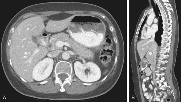
Imaging and Radiological Assessment of the Inferior Vena Cava | Thoracic Key

Trunk
Trunk

A) Chest CT scan shows superior vena cava (SVC) thrombosis until the... |  Download Scientific Diagram
A) Chest CT scan shows superior vena cava (SVC) thrombosis until the... | Download Scientific Diagram
File:Doppelte Vena cava inferior27jm - CT axial und coronar - 001 -  colored.png - Wikimedia Commons
File:Doppelte Vena cava inferior27jm - CT axial und coronar - 001 - colored.png - Wikimedia Commons

CT thorax visualizing persistent left superior vena cav | Open-i

Benign Central Superior Vena Cava Obstruction (17.09.2021)

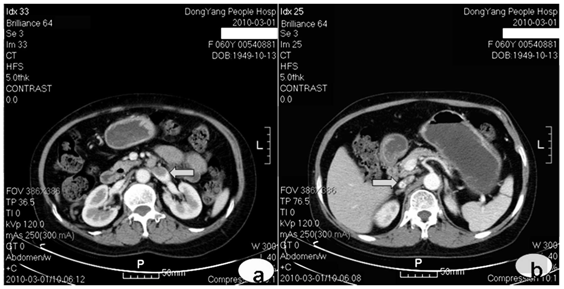
Computed tomography manifestations of common inferior vena cava dysplasia  and its clinical significance
Computed tomography manifestations of common inferior vena cava dysplasia and its clinical significance

Anomalous development of the inferior vena cava: Case reports of agenesis  and hypoplasia - ScienceDirect
Anomalous development of the inferior vena cava: Case reports of agenesis and hypoplasia - ScienceDirect

Axial post-contrast CT scan shows left lobe mass, inferior vena cava... |  Download Scientific Diagram
Axial post-contrast CT scan shows left lobe mass, inferior vena cava... | Download Scientific Diagram

Axial CT scan of the abdomen. A= Aorta, RV= Right Vena cava, LV= Left... |  Download Scientific Diagram
Axial CT scan of the abdomen. A= Aorta, RV= Right Vena cava, LV= Left... | Download Scientific Diagram

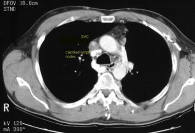
Superior Vena Cava Syndrome Imaging: Practice Essentials, Imaging Modalities

JCDD | Free Full-Text | A Unique Case of Inferior Vena Cava Aneurysm  Complicated with Pulmonary Embolism and Cerebral Infarction
JCDD | Free Full-Text | A Unique Case of Inferior Vena Cava Aneurysm Complicated with Pulmonary Embolism and Cerebral Infarction

Superior Vena Cava Obstruction Evaluation With MDCT | AJR

Two cases of inferior vena cava duplication with their CT findings and a  review of the literature. | Semantic Scholar
Two cases of inferior vena cava duplication with their CT findings and a review of the literature. | Semantic Scholar

The inferior vena cava: anatomical variants and acquired pathologies |  Insights into Imaging | Full Text
The inferior vena cava: anatomical variants and acquired pathologies | Insights into Imaging | Full Text
Double inferior vena cava mimicking lymphadenopathy | Annals of Saudi  Medicine
Double inferior vena cava mimicking lymphadenopathy | Annals of Saudi Medicine

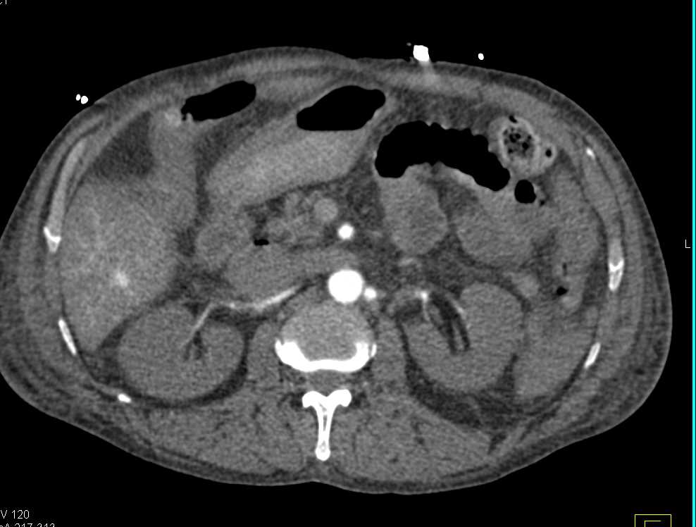
Dilated Inferior Vena Cava (IVC) with Poor Cardiac and Renal Function -  Liver Case Studies - CTisus CT Scanning
Dilated Inferior Vena Cava (IVC) with Poor Cardiac and Renal Function - Liver Case Studies - CTisus CT Scanning
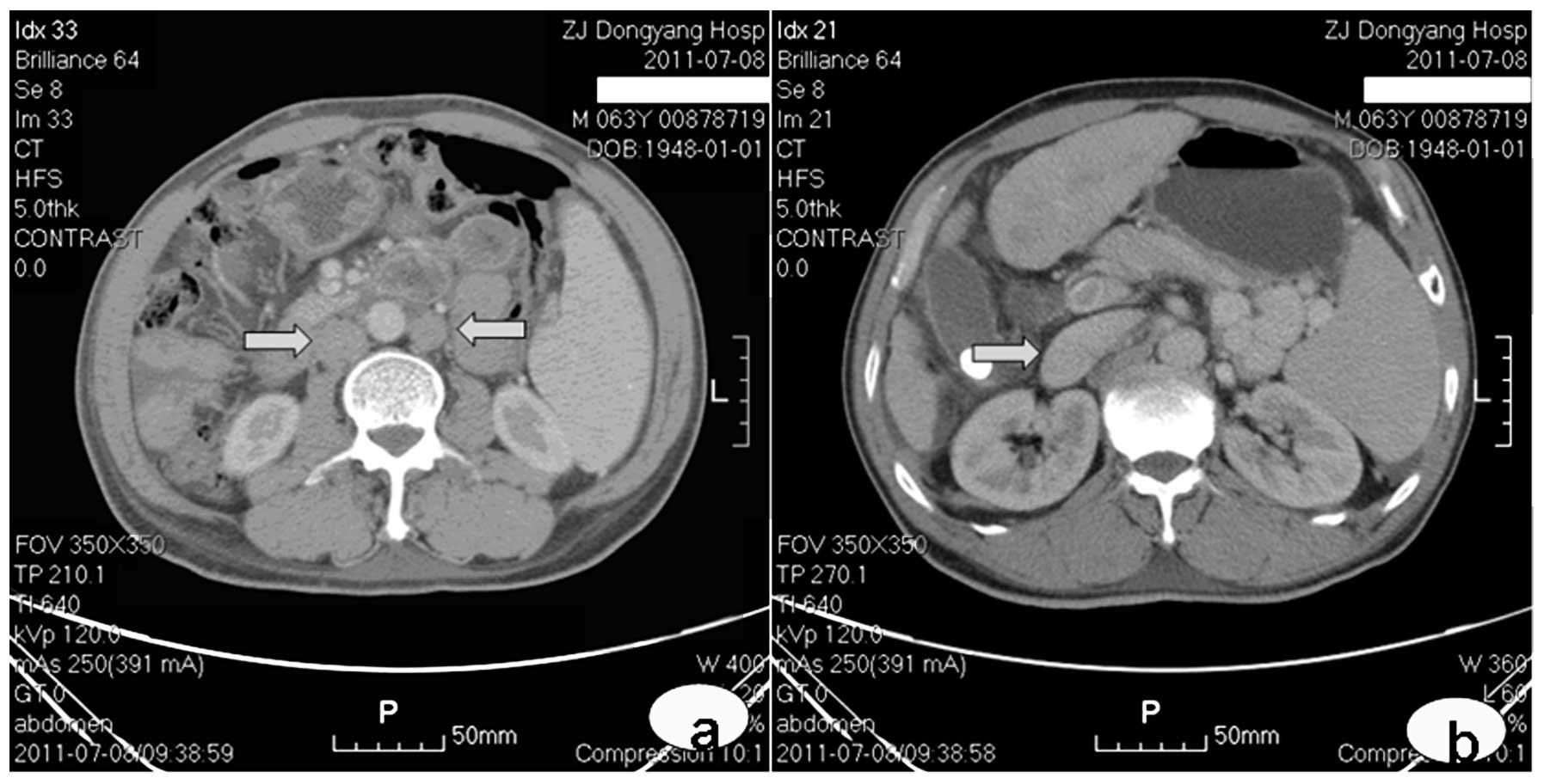
Computed tomography manifestations of common inferior vena cava dysplasia  and its clinical significance
Computed tomography manifestations of common inferior vena cava dysplasia and its clinical significance

CT abdomen. 1. Abdominal aorta 2: Inferior vena cava 3: Liver 4:... |  Download Scientific Diagram
CT abdomen. 1. Abdominal aorta 2: Inferior vena cava 3: Liver 4:... | Download Scientific Diagram

Abdominal CT: abdominal veins • LITFL • Radiology Library

Inferior Vena Cava and Its Main Tributaries | Radiology Key

Inferior vena cava | Radiology Reference Article | Radiopaedia.org

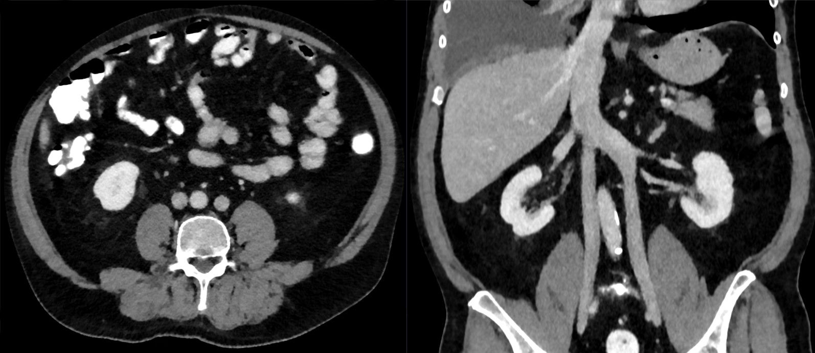
File:Doppelte Vena cava inferior 55M - CT axial paracoronar KM pv - 001.jpg  - Wikimedia Commons
File:Doppelte Vena cava inferior 55M - CT axial paracoronar KM pv - 001.jpg - Wikimedia Commons